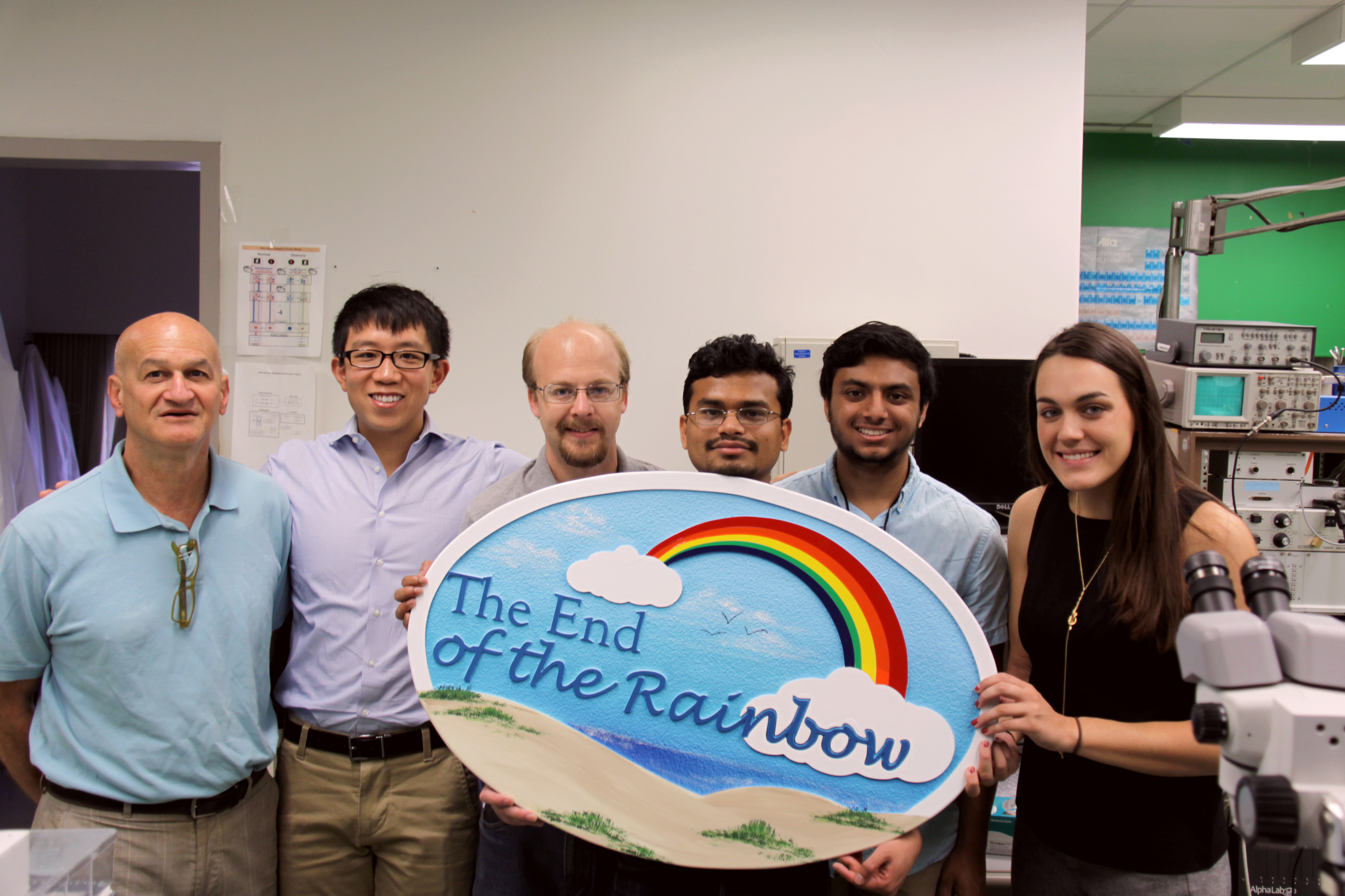
Pictured above is Dr. Mark Baron (far left) and his team of lab researchers.
Bench Research
Dystonia
Dystonia is the third most common movement disorder. It is a devastating condition characterized by ineffective, twisting movements and contorted postures. Dystonia prevents normal movements, causing instead twisted, distorted attempts to move. In the most severe cases, the afflicted is chair, and even bed, bound. Primary (genetic or idiopathic) dystonia manifests typically by adolescence and usually responds in part to therapeutics, with potential dramatic benefits from surgical intervention. Secondary (acquired) dystonia results from such causes as traumatic brain injury (TBI) or head trauma, stroke or cerebral palsy. In distinction from most primary dystonias, treatments for most secondary dystonias are largely ineffective. To date, the underlying abnormalities in the brain responsible for secondary dystonia have in particular had not been well studied. Consequently, secondary dystonia and its mechanisms are an important target for potentially life-changing research.
Secondary Dystonia Research
Dr. Mark Baron’s laboratory at the Veterans Affairs Medical Center has been devoted to understanding the underlying abnormal brain cell signaling responsible for dystonia and other movement disorders in animal models. His investigations in rodent models of dystonia have led to remarkable insights into how abnormal signals originating in the dystonic brain’s basal ganglia cause the thalamus to send pathological signals onto the motor cortex at the surface of the brain. These abnormal brain signals, in turn, ultimately lead to erroneous signals being sent to the muscles, leading to the devastating motor features of this condition.
Baron and his team discovered that the neuronal (brain cell) activity in a specific part of the basal ganglia, the globus pallidus (GP) in rodents (referred to in humans as the GPe) goes abnormally silent during dystonic movements in animals with experimental dystonia. To simulate the abnormal silencing of these neurons more permanently, the team next creates small destructive chemical lesions in GP in additional animals. As predicted, this caused secondary dystonia, which could be produced by destroying a specific identified tiny hotspot region of GPe. The researchers also discovered that destruction of an adjacent nearby separate hotspot region of GPe caused instead signs of Parkinson’s disease. This next led the team to discover that these same anatomical relations hold in humans undergoing deep brain stimulation; thereby, guiding improved targeting of deep brain stimulation for these two conditions. This work was published in Neuroscience (Kumbhare et al., 2017). Most recently, the team injected fluorescent-tagged viruses (with the ability to travel across connecting brain regions) into each of the hotspot regions of GP and traced these viruses to the surface of the brain. This revealed that in dystonia, the primary motor cortex (responsible for programming movement) receives the abnormal signals from the basal ganglia leading to the abnormal twisting movements. In the case of Parkinson’s disease, the abnormal signals are transmitted to the supplementary motor area (SMA) of the motor cortex, which is responsible for higher-level control of movement. Therefore abnormal signaling in the SMA in Parkinson’s disease prevents the normal signaling required to initiate and influence ongoing movements, leading to the abnormal features characteristic of this condition.
Building on the team’s successes, Dr. Baron and colleagues will soon begin investigating the utility of transcranial magnetic stimulation (TMS) over the skulls of the animals while targeting the newly identified underlying problematic brain regions (the primary motor cortex for Parkinson’s disease and the SMA for dystonia). With a further interest in understanding the underlying mechanisms and with additional potential therapeutic implications, the team will also be inserting opsins (proteins found in microorganisms) directly into brain cells and then subject these opsins to highly specific light waves with a goal to precisely program the abnormal brain cells to approach more natural signaling activity. Dr. Baron’s studies provide new hope to patients suffering from secondary and other forms of dystonia and for those with Parkinson’s disease and other disabling movement disorders.